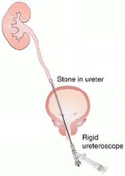
Ureteroscopy is where a long thin rigid telescope is introduced into the upper urinary tract via the bladder. The diameter of the instrument is less than 3 mm and allows visualisation of the lower half of the ureter. A small instrument port allows introduction of micro-baskets and laser fibres (0.3 mm in diameter) to manipulate and fragment stones. It is used only to treat stones in the ureter, and cannot treat stones within the kidney (see flexible pyeloscopy info sheet for kidney stone treatment.
What are the main advantages of this approach?
Allows stone treatment without the need for any incision by using the urethral orifice as the entry point
A highly successful technique (over 95%)
Can be performed as day surgery
What are the main disadvantages?
More invasive compared to shock wave lithotripsy
Small risk of damaging the ureter (0.5%)
What preparation is required?
As the procedure is performed under general anaesthesia, you should have nothing to eat or drink for 6 hours prior to treatment. Regular medications can be taken with a sip of water with the exception of blood thinning agents (eg. warfarin, aspirin, clopidogrel) or non-steroidal anti-inflammatories which need to be stopped for 7-10 days. A mid stream urine (MSU) test is required to ensure the urine is sterile before treatment is undertaken.
What do I need to bring to surgery?
All related imaging such as x-rays, CT scan or ultrasound
Your usual medications
What happens in the operating room?
You will meet your anaesthetist prior to surgery who will take a thorough medical history. This person will be responsible for your safety whilst you are under general anaesthesia. The procedure will usually take 60 minutes and involves putting a rigid telescope into the drainage tube (ureter) of the kidney and fragmenting the stone with laser. A temporary urinary stent may be left in place for a short period to ensure the kidney drains without risk of blockage.
What are the risks?
This is generally considered a very safe operation.
Specific risks to surgery include:
infection, minor bleeding, and perforation of the ureter (1 in 200).
What to expect afterwards?
It is normal to feel the need to pass urine frequently and notice blood in the urine following surgery. This will settle over the ensuing days. An oral over the counter medication called Ural can reduce the stinging sensation during urination. You will sometimes have a temporary urinary stent (see urinary tract stent info sheet) following surgery which allows the swelling in the ureter to settle from where the stone was located. The stent maybe attached to a string coming out from the urethra allowing ease of removal (in the doctor’s office) when no longer required. Care needs to be taken so as not to accidently dislodge the stent by pulling on the string or catching it on your underwear.
Follow-up
You will be advised after surgery the necessary follow-up arrangements. A script for oral antibiotics will need to be taken for 5 days to prevent infection. You need to drink at least 8 glasses of water a day (2.5L/day). Simple analgesics such as Panadol and Nurofen are usually all that is required, occasionally stronger medication (eg. Panadeine Forte) may be necessary. You will not be able to drive for at least 24 hours after surgery as you have had a general anaesthetic.
2. Laser Stone Surgery using Flexible Pyeloscopy
What is it?
Pyeloscopy is where a thin fibre-optic telescope is introduced into the kidney from the bladder via the urethra (see diagram). The diameter of the instrument is less than 3mm and allows visualisation of the entire kidney drainage system due to the flexible nature of the scope. It contains a small instrument port which allows the introduction of laser fibres (0.3 mm diameter) to efficiently fragment stones, and micro-baskets (less than 1mm wide) to retrieve stone fragments. Kidney stones up to 2 cm in size can be treated using this approach.
What are the main advantages of this approach?
Allows stone treatment without the need for any incision by using the urethra as the entry point Has a very high degree of success in treating stones (over 90-95%) Can be performed as day surgery
What are the main disadvantages?
More invasive compared with shock wave lithotripsy
General anaesthetic required
Small risk of damage to ureter (0.5%)
What preparation is required?
As the procedure is performed under general anaesthesia, you should have nothing to eat or drink for 6 hours prior to treatment. Regular medications can be taken with a sip of water with the exception of blood thinning agents (eg. warfarin, aspirin, clopidogrel) or non-steroidal anti-inflammatories which need to be stopped for 7-10 days. A mid stream urine (MSU) test is required to ensure the urine is sterile before treatment is undertaken.
What do I need to bring to surgery?
All related imaging such as x-rays, CT scan or ultrasound
Your usual medications
What happens in the operating room?
You will meet your anaesthetist prior to surgery who will take a thorough medical history. This person will be responsible for your safety whilst you are under general anaesthesia. The procedure will usually take 60 to 90 minutes and involves putting a flexible telescope into the drainage tube of the kidney and fragmenting the stone(s) with laser. A temporary urinary stent may be left in place for a short period to ensure the kidney drains without risk of blockage.
What are the risks?
This is generally considered a very safe operation.
Specific risks to surgery include:
infection, minor bleeding, and perforation of the ureter (1 in 200).
What to expect afterwards?
It is normal to feel the need to pass urine frequently and notice blood in the urine following surgery. This will settle over the ensuing days. An oral over the counter medication called Ural can reduce the stinging sensation during urination. You will sometimes have a temporary urinary stent (see urinary tract stent info sheet) following surgery which allows the stone fragments to drain unimpeded. The stent maybe attached to a string coming out from the urethra allowing ease of removal (in the doctor’s office) when no longer required. Care needs to be taken so as not to accidently dislodge the stent by pulling on the string or catching it on your underwear.
Follow-up
You will be advised after surgery the necessary follow-up arrangements. A script for oral antibiotics will need to be taken for 5 days to prevent infection. You need to drink at least 8 glasses of water a day (2.5L/day). Simple analgesics such as Panadol and Nurofen are usually all that is required, occasionally stronger medication (eg. Panadeine Forte) may be necessary. You will not be able to drive for at least 24 hours after surgery as you have had a general anaesthetic.
3. Percutaneous Nephrolithotomy (PCNL)
What is it?
Percutaneous Nephrolithotomy (PCNL) is the preferred technique for treating large stones (over 2 cm in diameter) within the kidney. It involves keyhole surgery performed through a 1cm incision in the skin overlying the kidney (see diagram).
What are the advantages?
Allows large or complicated stones to be treated in a minimally invasive fashion, where in the past this would have necessitated a large skin incision.
Hospital stay is 3-4 days, and out of hospital recovery time is significantly shorter than traditional open surgery.
What are the disadvantages?
Compared to traditional treatments of large complex stones, there are no disadvantages. Improved techniques and equipment have allowed this type of surgery to be safer than ever before.
What preparation is required?
You will be required to have detailed imaging to allow the surgeon to assess the stone in fine detail regarding its relationship to the kidney and nearby structures. This will enable the surgeon to plan the best access point(s) to the kidney to allow effective clearance of stones. As the procedure is performed under general anaesthesia, you should have nothing to eat or drink for 6 hours prior to treatment. Regular medications can be taken with a sip of water with the exception of blood thinning agents (eg. warfarin, aspirin, clopidogrel) or non-steroidal anti-inflammatories which need to be stopped for 7-10 days. A mid stream urine (MSU) test is required to ensure the urine is sterile before treatment is undertaken. Other tests required include urine culture, kidney function studies, and complete blood counts. These tests will all be organized from the rooms after your consultation.
What are the risks?
This form of surgery is low risk if performed by an urologist who is specifically trained in this technique, and aided by meticulous pre-operative planning. The specific risks are uncommon but include infection, excessive bleeding (necessitating blood transfusion 2%, embolisation 1%, renal exploration 0.5%), and adjacent organ injury (spleen, liver, bowel, and lung).
What do I need to bring to surgery?
All related available imaging such as KUB (kidneys, ureter, and bladder) x-ray, CT scan abdomen, or kidney ultrasound
Your usual medications
What happens in the operating room?
The operation is performed under a general anaesthetic and lasts approximately 2 to 3 hours. It is a team effort requiring coordination between surgeon, anaesthetist, radiology and nursing staff. You will be positioned on the operating room table lying on your front “stomach” for the duration of the surgery. The procedure is accomplished with the assistance of x-ray imaging to guide entry of a 1 cm tube into the kidney. This provides access into the kidney drainage system allowing telescopes and instruments to visualize, fragment and remove stones. A drainage catheter (nephrostomy tube) which exits through the skin is left in the kidney at the end of the procedure.
What to expect afterwards?
You will have a temporary catheter called a nephrostomy tube draining the kidney, as well as a urinary catheter in-situ. They will be removed prior to discharge from hospital. The urine will be bloodstained for up to a week after discharge from hospital. Imaging is performed immediately after surgery to assess stone clearance. Occasionally, further minor surgery is required to clear any remaining stones to achieve complete stone clearance. Your hospital stay will be 3-4 days on average.
Follow-up
You will be required to take it easy during the recovery phase for several weeks. There should only be minimal discomfort from the wound. Oral antibiotics will be given for a further five days to prevent infection. It is important to inform us if you feel unwell with fevers, chills, or develop heavy bleeding in the urine. Your initial follow-up will be in 6 weeks after discharge. Occasionally, a urinary stent is left to ensure the urine drains correctly into the bladder. This will require removal at a later time.
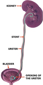
4. Urinary Tract "Double J" Stent
What is it?
A thin, hollow tube placed inside the ureter during surgery to ensure drainage of urine from the kidney into the bladder. J shaped curls are present at both ends to hold the tube in place and prevent migration, hence the description "Double J stent".
Purpose?
It allows the kidney(s) to drain urine by temporarily relieving any blockage, or to assist the kidney(s) in draining stone fragments freely into the bladder if definitive kidney stone surgery is carried out.
What symptoms will you notice?
This tube has curls at either end which keeps the stent from moving. The lower curl can irritate the bladder and make you feel the need to go to the toilet all the time to empty your bladder. It can also cause moderate discomfort in the kidney region on urination, and at times you may notice blood in the urine. None of these symptoms are harmful and your body gets use to the stent over time. Simple analgesics may be helpful in controlling the discomfort (Panadol or Nurofen). All the symptoms resolve after stent removal once definitive treatment is completed.
What if there is a string present?
Sometimes there is a string attached to the stent coming from your urinary passage and taped to the nearby skin. Your surgeon will let you know if this occurs after your surgery. It is often performed when definitive stone treatment is carried out and allows the stone fragments to drain out freely from the kidney. This is for your benefit as it allows stent removal in our office by simply pulling on the string (7 days) without the need for further surgery. You will be advised when it can be taken out. If you accidentally dislodge the stent, you will notice being constantly incontinent of urine. If this occurs, you can safely remove the stent by pulling on the string. Call the office the next day to inform us.
Important Information!
Stents are used in temporary situations and must be removed from the body. Occasionally, some events occur where communication and follow up is lost and you must be aware that these stents should be removed within six (6) months from the date of insertion. In most cases they will be removed well before that time. It is important that you remember you have a stent and not forget about it.

 What is it?
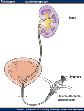 2. Laser Stone Surgery using Flexible Pyeloscopy
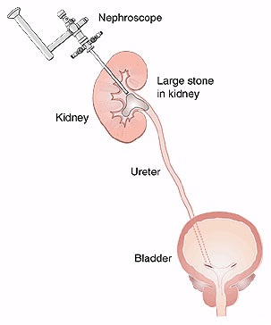 What are the advantages?